h1.qusth1 { display: none !important; }

QUESTION OF THE WEEK

Dr. Donovan's Articles

QUESTION OF HAIR BLOGS


OMAHA EFFECT? Oral Minoxidil Alcohol Hangover Association

Oral Minoxidil Might Increase the Possibility of Alcohol Hangover.

If you are like me and never heard of the OMAHA effect, then read on!

Oral minoxidil is increasingly used for the treatment of hair loss. Authors from New York recently reported ten patients who used oral minoxidil and developed alcohol hangover symptoms with alcohol. These symptoms included nausea, headaches, and light sensitivity.


The authors note that an online interaction checker showed that alcohol and minoxidil do have a moderate interaction – because the two have an added effect on lowering blood pressure. I noted that when I checked drugs.com today, this same effect could be seen.


In this paper, the authors define an alcohol hangover as “a combination of negative mental and physical symptoms which can be experienced after a single episode of alcohol consumption, starting when blood alcohol concentration (BAC) approaches zero.”  The authors remind us that symptoms of alcohol hangover might include headache, nausea, fatigue, dry mouth, third and may encompass fatigue, headaches, increased thirst, and dry mouth.

The authors point out that the exact mechanisms still need to be clarified. Still, there are suggestions in animal studies that oral minoxidil could affect how alcohol is metabolized in the gastrointestinal tract. Overall, the authors point out that this is not something that is seen with other blood pressure drugs. Specifically, they state that there have been no reports of other blood pressure-lowering drugs - particularly those with vasodilatory properties such as hydralazine or calcium channel blockers causing any exacerbating alcohol hangover symptoms.

The authors state that in their practice, they advise patients to refrain from taking their LDOM dose the day they anticipate excessive alcohol consumption. And they call for further research on the potential worsening of alcohol hangovers with oral minoxidil.

Why this study caught my attention?

This study caught my attention for two reasons. First, this is an interesting observation. Having been prescribed oral minoxidil for a long time, I’ve not seen or heard of this before. I’m going to keep watch and start asking patients.

Second, I’m surprised this article was accepted for publication without requiring more information. While the article reads beautifully, there is no quantitative information in this paper except for the number of patients the authors have seen report this effect. We don’t have information on who these patients are, what doses they took, what symptoms of alcohol hangover they had, how long the symptoms lasted, or how sick patients were. I’ve reread the paper multiple times to see if I missed it. I didn’t - there is nothing in here. I think new, potentially practice-changing observations require that journal editors demand more.

Nevertheless, you can be assured that I’ll consider the OMAHA effect when I see my patients today.

REFERENCE

Lina Alhanshali 1, Deesha Desai 2 3, Ambika Nohria 3, Jerry Shapiro 3, Kristen Lo Sicco 3. Oral minoxidil and the exacerbation of alcohol hangover symptoms. Affiliations expanInt J Dermatol . 2024 Apr 21. doi: 10.1111/ijd.17195. Online ahead of print.


This article was written by Dr. Jeff Donovan, a Canadian and US board certified dermatologist specializing exclusively in hair loss.
Share This
No Comments

Most practitioner - but not all - think oral minoxidil

I enjoyed reading a study by Sanfilippo and Friedman in the March issue of the Journal of Drugs in Dermatology about practitioners' opinions on the benefits of oral minoxidil.

Oral minoxidil is increasingly popular. What matters most to me is what the science says. Is oral minoxidil helpful, and is it better than topical minoxidil, and what are the long-term side effects of oral minoxidil after 35 years of use? It’s now clear that oral minoxidil is helpful. It’s not entirely clear if it’s better than topical minoxidil - at least based on wonderful studies like the study we reviewed by Penha et al. That study showed that oral minoxidil failed to beat topical minoxidil in hair growth. The two were somewhat similar. However, just when you think the case is sealed - and you are about to write in the history books forever that oral minoxidil and topical minoxidil are equivalent - those same authors showed that photographic assessments done by doctors suggested that oral minoxidil was, in fact, better than topical minoxidil for helping hair growth in the crown but pretty equivalent for helping improve hair in the frontal scalp.

If I spent my days simply reviewing the most incredibly well-conducted studies in the world, I’d miss out on a lot of important information. What matters to me is what patients think about specific diseases and treatments and what healthcare practitioners feel about specific diseases and treatments. I call these types of studies the “pulse” of the world!

I’m interested in this information not because it influences WHAT I do in my clinic but because it influences HOW I do it. If repeated studies show convincingly that drug A is better than drug B, but repeated surveys show that the entire planet (patients and doctors alike) still feels drug B is better than drug A, does it mean that I proceed to prescribe drug B? Well, no, but it means that I have my work cut out to counsel patients, and I have my work cut out to explain things to my colleagues.

The Sanfilippo and Friedman study of 2024 is one of those studies that tell me what sort of work I have waiting when discussing oral minoxidil with colleagues. The study showed that approximately 10 % of practitioners don’t feel comfortable prescribing oral minoxidil, 10 % don’t feel oral minoxidil is well tolerated by patients, approximately 20 % don’t feel oral minoxidil is better than topical, and 20 % feel their patients aren’t so satisfied with results.

Now, this is not a study of who is right and who is wrong—it is an estimate of practitioners' views. This is the pulse!


Sanfilippo and Friedman 2024


The authors set out to evaluate dermatology providers' attitudes and recommendation behaviours of oral minoxidil for treating AGA. They performed an online survey. The survey was sent to those attending the Orlando Dermatology Aesthetic and Clinical Conference. The survey participants included many types of dermatology practitioners, including MD/DOs, NPs, and PAs across the United States.

What were the results?

The survey was sent to about 2,200 providers. The response rate was poor, and just 201 (9.1%) of surveys came back. This, of course, opens the door to all sorts of bias. People who love oral minoxidil might be more likely to fill out the survey…. or maybe people who detest oral minoxidil might be more likely to fill out the survey. Who knows?

Overall, here are some key points:

a) 81% (n=139) of respondents supported using oral minoxidil for AGA.

b) Those in practice over 30 years gave the least support.

c) 92% of respondents stated they were comfortable prescribing oral minoxidil

d) 83% of respondents felt oral minoxidil was more effective than its topical formulation.

e) 78% of respondents felt their patients were satisfied with the results of using oral minoxidil

f) 89% of respondents felt oral minoxidil was well tolerated by their patients.


Comments

I liked this study. Despite the extremely low response rates, this study has much to offer. It’s clear that positive views outweigh the negative ones - but the negative ones should not be discarded. Not everyone shares the same love and infatuation for oral minoxidil. Not every one of our colleagues has set sail on the oral minoxidil cruise ship.

Let’s face it—at least in this study—10 % of practitioners don’t feel comfortable prescribing oral minoxidil, 10 % don’t feel it is well tolerated by patients, approximately 20 % don’t feel it is better than topical, and 20 % feel their patients aren’t so satisfied with results.

If you believe that long-time practitioners are wise and experienced, you’ll find it interesting that they are less likely to be in love with oral minoxidil.

If you believe that long-time practitioners are inflexible, rigid in their ways, and unwilling to try new things, then you may not be surprised that this group is less likely to be in love with oral minoxidil.

What’s your view?

Congratulations to these authors for a wonderful study highlighting the “pulse” of oral minoxidil amongst practitioners in the United States.

REFERENCES

Eric Sanfilippo and Adam Friedman. Survey of Dermatology Practitioners' Opinions and Prescribing Habits of Oral Minoxidil for the Treatment of Androgenetic Alopecia. J Drugs Dermatol . 2024 Mar 1;23(3):136-140. doi: 10.36849/jdd.7519.


Mariana Alvares Penha 1, Hélio Amante Miot 1, Michal Kasprzak 2, Paulo Müller Ramos. Oral Minoxidil vs Topical Minoxidil for Male Androgenetic Alopecia: A Randomized Clinical Trial. JAMA Dermatol . 2024 Apr 10:e240284. doi: 10.1001/jamadermatol.2024.0284. Online ahead of print


This article was written by Dr. Jeff Donovan, a Canadian and US board certified dermatologist specializing exclusively in hair loss.
Share This
No Comments

Alopecia Areata Linked to Pollen, Dust, and Cat Allergies

Authors from China recently performed a large study to assess the relationship between alopecia areata and various allergic conditions, including hay fever, eczema, asthma, and allergies to pollen, dust, and cats.

Interestingly, the authors’ data showed a significant genetic correlation between atopy/allergies and an increased risk of developing alopecia. Notably, strong associations were observed for eczema, hay fever, asthma, and specific allergies to pollen, dust, and cats. All these conditions are associated with an increased risk to develop alopecia areata.

The authors conclude that there is compelling genetic evidence of an association between atopic and allergic conditions and the development of alopecia areata.

REFERENCE

Xu W et al. Genetic links between atopy, allergy, and alopecia areata: insights from a Mendelian randomization study. Allergy Asthma Clin Immunol. 2024 Apr 27;20(1):32. doi: 10.1186/s13223-024-00892-w.


This article was written by Dr. Jeff Donovan, a Canadian and US board certified dermatologist specializing exclusively in hair loss.
Share This
No Comments

Air Pollutants are a Likely Contributor to The Rising Incidence of Autoimmune Diseases

I’m amazed at the number of studies I read nowadays linking air pollution, water pollution and climate change in general to the rise in autoimmune diseases.

Wen et al 2024

A study by Wen et al investigated the possible role of air pollution in 15 autoimmune diseases. Data from the authors revealed that hypothyroidism, ulcerative colitis, rheumatoid arthritis, systemic lupus and celiac disease were all among the group of diseases causally associated with higher exposure to air pollution.

The authors propose that their research underscores the necessity of rigorous air pollutant surveillance within public health studies to curb the prevalence of autoimmune diseases.

Hu et al 2024

A new study by Hu et al. showed an association between small particulate matter and the risks of psoriasis, vitiligo, and lupus in a European population.

REFERENCES

Wen J et al. Large-scale genome-wide association studies reveal the genetic causal etiology between air pollutants and autoimmune diseases. J Transl Med . 2024 Apr 29;22(1):392. doi: 10.1186/s12967-024-04928-y

Hu H et al. Causal association between air pollution and autoimmune diseases: a two-sample Mendelian randomization study. Front Public Health . 2024 Mar 28:12:1333811. doi: 10.3389/fpubh.2024.1333811. eCollection 2024.


This article was written by Dr. Jeff Donovan, a Canadian and US board certified dermatologist specializing exclusively in hair loss.
Share This
No Comments

Dutasteride May Reduce Heavy Drinking in Males

5-alpha reductase enzymes have an important role in the production of 5α-reduced neuroactive steroids. These molecules may affect human drinking behaviours and alcohol consumption. Authors of a new study set out to evaluate dutasteride's tolerability and efficacy for reducing drinking.

The study included 142 males with heavy drinking (defined as more than 24 drinks per week) and a goal to reduce or stop their drinking were randomized to either dutasteride 1 mg daily or placebo for 12 weeks.

Results showed that 25% of dutasteride-treated participants had no hazardous drinking (no heavy drinking days and not more than 14 drinks per week) during their last month of treatment compared with 6% of placebo-treated participants (P = 0.006; NNT = 6). Adverse events more common in the dutasteride group included reduced libido and stomach discomfort.

Conclusion:

This study positions dutasteride 1 mg was a medication to reduce heavy drinking in males. More studies are needed to understand the long term effects of dutasteride on drinking behaviours.

REFERENCE


Covault J et al. Randomized Placebo-Controlled Clinical Trial of Dutasteride for Reducing Heavy Drinking in Men. J Clin Psychopharmacol. 2024 May-Jun;44(3):223-231. doi: 10.1097/JCP.0000000000001849.


This article was written by Dr. Jeff Donovan, a Canadian and US board certified dermatologist specializing exclusively in hair loss.
Share This
No Comments

New study reveal iron deficiency and iron deficiency anemia is common in Canada

40 % of women aged 15-54 have low ferritin and 13 % are anemic

Low ferritin can sometimes contribute to hair shedding - especially as ferritin drops well below 40. Authors of an interesting new study set out to determine the prevalence of iron deficiency and iron deficiency anemia in women 15-54 in the province of Ontario Canada. Iron deficiency was defined as ferritin <30 μg/L. Anemia was defined as hemoglobin <120 g/L and Iron deficiency anemia as ferritin <30 μg/L and hemoglobin <120 g/L.

Over 700,000 women were evaluated in this study. Surprisingly, 38.3% of women had low ferritin, and, 13.1% had iron deficiency anemia; 55.6% of women with iron deficiency anemia still had normal mean corpuscular volumes.

COMMENT

All women with concerns about hair loss need basic blood tests like a complete blood count, ferritin level, thyroid stimulating hormone level and 25 hydroxvitamin D level. It’s incredible just how prevalent iron deficiency is in Canada.

REFERENCE


Wen S et al. High prevalence of iron deficiency and socioeconomic disparities in laboratory screening of non-pregnant females of reproductive age: A retrospective cohort study. Am J Hematol. 2024 May 2. doi: 10.1002/ajh.27352. Online ahead of print.


This article was written by Dr. Jeff Donovan, a Canadian and US board certified dermatologist specializing exclusively in hair loss.
Share This
No Comments

Oral Minoxidil Fails to Beat Topical Minoxidil in Improving Hair Counts

Is oral minoxidil more effective than topical minoxidil?


A new randomized controlled study suggests that male users of 5 mg oral minoxidil once daily do not seem to get all that much more hair growth on the scalp compared to those using 5 % topical minoxidil twice daily.


As you’re probably well aware, oral minoxidil was originally used as a blood pressure medication. In fact, the FDA approved it for the treatment of hypertension back in 1979. Typical blood pressure doses were 10 to 40 mg daily. At these doses, the most common adverse effects are hypertrichosis, tachycardia, headache, and edema. It soon became clear in the early 1980s that oral minoxidil is a treatment option for males with AGA. Topical minoxidil received FDA approval for male balding just 9 years later - in 1988.  


To date, there has been incredible interest in oral minoxidil. Surprisingly, despite this interest, only a few high-quality studies have been done. We discussed the Panchaprateep et al. study in the past. It wasn’t a super high-quality study, but it represented the best we had - until now.


As we’ll see now in the nicely conducted RCT by Penha et al, oral minoxidil did not seem to perform so well in males in Brazil. Specifically, it was unable to beat topical minoxidil in hair growth.


 Penha et al 2024

Authors from Brazil set out to compare the efficacy, safety, and tolerability of daily oral minoxidil, 5 mg, vs twice-daily topical minoxidil, 5%, for 24 weeks in the treatment of male AGA.


This was a double-blind, placebo-controlled, randomized clinical trial conducted at a single specialized clinic in Brazil. Eligible men with AGA aged 18 to 55 years classified using the Norwood-Hamilton scale as 3V, 4V, or 5V were included and randomized.  Participants were randomized 1:1 into 2 groups: oral minoxidil, 5 mg, daily and topical placebo solution; or 1 mL of topical minoxidil, 5%, twice daily and oral placebo for 24 weeks.


The oral minoxidil group received 5 mg of minoxidil capsules plus a placebo solution to apply to the scalp. The topical minoxidil group received a 5% topical minoxidil solution plus placebo capsules.


The primary outcome was the change in terminal hair density on the frontal and vertex regions of the scalp. Secondary outcomes included change in total hair density in the target area, assessment of standardized clinical photographs, and assessment of adverse effects, blood pressure, and heart rate.


Among 90 enrolled participants, 68 completed the study; of these, the mean age was about 37 years.  A total of 33 participants were enrolled in the oral minoxidil group and 35 in the topical treatment group. Both groups were homogenous in terms of demographic data and AGA severity.


Most participants had mild to moderate AGA, and the groups were homogeneous. A total of 68 completed the study and had 24-week follow-up data. There were 12 dropouts (27%) in the oral minoxidil group and 10 drop outs in the topical group.   There was no difference in the proportions of dropouts between groups.


When comparing oral minoxidil to topical minoxidil for the frontal area

·      After 24 weeks, males using oral minoxidil had 3.1 more terminal hairs per cm2 (95% CI, −18.2 to 21.5; P = .27) and 2.6 hairs per cm2 (95% CI, −10.3 to 15.8; P = .32) for total hair density.  None of this was statistically significant.


When comparing oral minoxidil to topical minoxidil for the vertex area

·      After 24 weeks, males using oral minoxidil had 23.4 more terminal hairs per cm2 (95% CI, −0.3 to 43.0; P = .09) and 5.5 hairs per cm2 (95% CI, −12.5 to 23.5; P = .32) for total hair density. None of this was statistically significant.


When examining “percent differences” instead of absolute differences, the authors found some differences.


FOR TERMINAL HAIR DENSITY CHANGES In the VERTEX, the percentage increase in terminal hair density was 27.1% (95% CI, 6.5-47.8) higher for the oral minoxidil group than the topical group (P = .005). This was statistically significant.


FOR TERMINAL HAIR DENSITY CHANGES In the FRONTAL SCALP, the percentage increase in terminal hair density was 13.1% higher for the oral minoxidil group (95%CI,−11.5 to 37.5%;P = .15). However, this was not statistically significant.


FOR TOTAL HAIR DENSITY IN THE VERTEX, there was an increase of 2.1% (95% CI, −8.1 to 12.3; P = .27) in the oral minoxidil group compared with the topical minoxidil group. This was not a statistically significant difference


FOR TOTAL HAIR DENSITY IN THE FRONTAL SCALP, there was a decrease of 0.2% (95% CI, −8.4 to 8.0; P = .89) ) in the oral minoxidil group compared with the topical minoxidil group. This was not a statistically significant difference.


PHOTOGRAPHIC ANALYSIS

According to the photographic analysis of the 3 dermatologists blinded to treatments, oral minoxidil was superior to topical minoxidil on the vertex (24%; 95% CI, 0 to 48; P = .04) but not on the frontal scalp (12%; 95% CI, −12 to 36; P = .24).

Specifically, 60% of males in the oral minoxidil group (60%) and 48% topical minoxidil group (48%) were rated to have a clinical improvement in the frontal area, with no significant difference between the groups (difference, 12%; 95% CI, −12 to 36;P = .24).  For the vertex, 70% of patients in the oral minoxidil were rated to have a clinical improvement in the vertex area compared to 46 % in the topical minoxidil group (16 of 35 [46%]) (difference, 24%; 95% CI, 0-48; P = .04). This was statistically significant.


SIDE EFFECTS

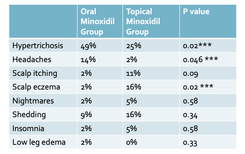
The most common adverse effects in the oral minoxidil group were hypertrichosis (22 of 45 [49%]) and headache (6 of 45 [14%]).  The most common side effect in the topical minoxidil group was hypertrichosis present in 25 % followed by scalp eczema in 16 % of users. There was no difference between the groups regarding variation in mean arterial blood pressure over time. Transient hair loss in the first two months was experienced by treatment by 9% in the oral minoxidil group and 16% in the topical minoxidil group, but this was not statistically significantly different.


COMMENT


This is an important study for the history books. First, it is the first well done comparative therapeutic trials of low-dose oral minoxidil for male AGA.


The study showed that oral minoxidil, 5 mg, once per day for 24 weeks did not demonstrate superiority over topical minoxidil, 5%, twice per day in men with AGA – at least for hair growth and the primary end point of actual numbers.


Despite these data, there is a hint that oral minoxidil is better for the crown in males. Expert photographic evaluation at the vertex and the percent increase of terminal hair density in the vertex area suggests that oral minoxidil probably beats out topical minoxidil.

Hypertrichosis was the main adverse effect reported and was more prevalent in the oral minoxidil group. Headache was more prevalent in the oral minoxidil group. Transient hair loss in the first two months was experienced by treatment by 9% in the oral minoxidil group and 16% in the topical minoxidil groups.

This study is a nice reminder of the true power of randomized trials. This study gave far inferior results for oral minoxidil than the much loved 2020 study by Panchaprateep et al. in the 2020 study, 93 % of oral minoxidil users had nice results compared to 69 % here in the Penha et al study. Further more, there was not a single user in the Panchaprateep et al. who failed to benefit from oral minoxidil. In contrast, about 1/3 of patients in the Penha et al study experienced no improvement or actually to worse.

The debate on exactly how well oral minoxidil works is far from over!


REFERENCE


Mariana Alvares Penha 1, Hélio Amante Miot 1, Michal Kasprzak 2, Paulo Müller Ramos. Oral Minoxidil vs Topical Minoxidil for Male Androgenetic Alopecia: A Randomized Clinical Trial. JAMA Dermatol . 2024 Apr 10:e240284. doi: 10.1001/jamadermatol.2024.0284. Online ahead of print


This article was written by Dr. Jeff Donovan, a Canadian and US board certified dermatologist specializing exclusively in hair loss.
Share This
No Comments

What Drugs Cause Alopecia Areata?

The authors of a new study set out to determine drugs implicated in the development of alopecia areata (so-called drug-induced AA). Ezemma et al. performed a systematic review using PubMed to search for all published cases of drug-induced AA.

The authors found sixty-six articles (55 case reports, 9 case series, and two retrospective studies) for their systematic review. These 66 articles included 102 patients. The average age was around 40 years, and approximately half of the patients were female (53.9%).

What drugs are implicated in Drug-Induced AA?

Thirty drugs were found to be associated with the development of drug-induced AA. Monoclonal antibodies caused seventy-six percent of drug-induced AA cases. The TNF-alpha inhibitors and dupilumab comprised most of these cases. Overall, TNF inhibitors were responsible for 47.0% of all patient cases and dupilumab 12.7%.

Adalimumab was the most common TNF inhibitor implicated in drug-induced AA. (18.6% of all cases). Other TNF inhibitors included infliximab (14.7%), etanercept (11.7%), and golimumab (1.9%)

The TOP Causes of Drug-Induced AA

Here is the list of top culprits in drug-induced AA:

  1. Adalimumab (18.6% of all published cases) - TNF inhibitor

  2. Infliximab (14.7% of all published cases) - TNF inhibitor

  3. Dupilumab (12.7% of all published cases) - anti-IL-4/13

  4. Etanercept (11.7% of all published cases) - TNF inhibitor

  5. Alemtuzumab (5.8% of all published cases) - monoclonal anti-CD52 antibody

  6. Hepatitis C therapy (ribavirin, interferon-alpha) (3.9% of all published cases)

  7. Cyclosporine (2.9% of all published cases) - calcineurin immunosuppressive agent

  8. Belimumab (2.9% of all published cases) - monoclonal AB; inhibitor of B-lymphocyte stimulator (BLyS)

  9. Cladribine (1.9% of all published cases) - antimetabolic chemotherapeutic

  10. Golimumab (1.9% of all published cases) - TNF inhibitor

  11. Haloperidol (1.9% of all published cases) - antipsychotic

  12. Pembrolizumab (1.9% of all published cases) - checkpoint inhibitor, anti-PD1

  13. Rifampicin (1.9% of all published cases) - antimicrobial

  14. Oral retinoid (1.9% of all published cases) - vitamin A derivative

Epidemiology of Drug-Induced AA

Overall, patchy AA was the most common presentation. This was seen in 70.5% of drug-induced AA cases.

The average time to onset of AA was 11.7 months, so this was not always seen immediately.

Interestingly, most patients had no family history of AA (93.1%).

What is the effect of stopping the culprit drug?

Forty-eight patients discontinued the drug thought to cause AA, and nearly all patients had regrowth regardless of additional treatment.

What if patients chose not to stop?

Forty-two patients continued the drug, and most did not have regrowth unless they underwent additional treatment. However, the vast majority (75%) of patients who continued the implicated drug and received treatment had hair regrowth.

Comment

This is interesting and provides some beneficial information. The implicated drugs are similar to those reported in Ravipati et al. (see link) but not the same. The most common drug in Ravipati is the taxanes.

REFERENCE*

Ezemma O et al. Drug-induced alopecia areata: A systematic review. J Am Acad Dermatol. 2024 Jan;90(1):133-134. doi: 10.1016/j.jaad.2023.05.022. Epub 2023 May 18.


This article was written by Dr. Jeff Donovan, a Canadian and US board certified dermatologist specializing exclusively in hair loss.
Share This
No Comments

Drug Induced Alopecia Areata : A Closer Look at the FAERS Database

Introduction

Medications have the potential to trigger many diseases. This includes hair loss. It’s always important to pay close attention to drugs that cause or trigger a specific disease because this provides important clues to the underlying mechanisms governing the disease pathogenesis.

The FDA Adverse Events Reporting System (FAERS): A Powerful Tool to Track Post-Marketing Side Effects

The Food and Drug Administration (FDA) launched a publicly available database that allows individual patients, healthcare professionals, and manufacturers to report adverse events caused by medications.

This data is essential to better understanding “real-world” side effects. By real-world, I mean side effects outside a well-controlled, highly monitored clinical trial.

There are now over 26 million side effects reported on the FDA database.

Ravipati A et al, 2024

The authors of a new study set out to review the FAERS database to understand better medications associated with alopecia areata.

The authors found 1,331 AA cases reported on FAERS since 1997. Reports are increasing over time. Approximately 75.6% of AA reports were in females, whereas 24.4% were in males. Most cases were in patients ages 18-64, but there are pediatric patients in the database and patients over 65.

Top 10 Medications Associated with AA in FAERS

The following are the ten medications:

  1. 29.2% of AA cases were seen with docetaxel (a taxane chemotherapy drug)

  2. 8.3% were with adalimumab (a tumour necrosis factor (TNF) inhibitor)

  3. 4.7% with trastuzumab (monoclonal antibody; HER2 inhibitor cancer therapy)

  4. 4.4% with cyclophosphamide (alkylating agent- used in cancer treatment and autoimmune dz)

  5. 4.3% with infliximab (a tumour necrosis factor (TNF) inhibitor)

  6. 4.1% with carboplatin (alkylating agent- used in cancer treatment

  7. 3.4% with dupilumab (monoclonal antibody, blocks IL4 and IL13)

  8. 2.7% with levonorgestrel (progestin)

  9. 2.6% with etanercept (a tumour necrosis factor (TNF) inhibitor)

  10. 2.6% with from pertuzumab (monoclonal antibody; HER2 inhibitor cancer therapy)

A Closer Look at the Top 10

Monoclonal antibodies accounted for 6 out of the top 10 drugs. 3 of the top 10 medications were chemotherapeutic agents, including two HER2 receptor inhibitors and carboplatin.

Among the monoclonal antibodies, males were more likely to report AA when taking adalimumab (OR: 1.79, P = 0.04) and dupilumab (OR: 2.56, P = 0.03).

Other less common reported causes in the FAERS (rank order 11-30)

11. methotrexate (antimetabolite used in cancer therapy and autoimmune disease)

12. levocetirizine (antihistamine)

13. secukinumab (interleukin-17A (IL-17A) receptor inhibitor)

14. montelukast (leukotriene receptor antagonist - used in allergy treatment)

15. Olopatadine (selective histamine H1 antagonist and mast cell stabilizer)

16. prednisolone (corticosteroid)

17. tacrolimus (calcineurin inhibitor immunosuppressant)

18. doxorubicin (anthracycline chemotherapeutic drug)

19. fexofenadine (antihistamine)

20. tofacitinib (JAK 1/3 inhibitor)

21. levothyroxine (thyroid medication)

22. azathioprine (antiproliferative and immunosuppressive agent)

23. ustekinumab (monoclonal antibody, anti IL-12/23 inhibitor)

24. sertraline (selective serotonin reuptake inhibitor (SSRI)

25. simvastatin (cholesterol medication)

26. leflunomide (disease-modifying anti-rheumatic drug)

27. paclitaxel (taxane chemotherapeutic)

28. dexamethasone (corticosteroid)

29. certolizumab (a tumour necrosis factor (TNF) inhibitor)

30. Ribavirin (nucleoside analogue, used in hepatitis C treatment)

Comment

This is an interesting paper that highlights the new world of drug-induced AA. Monoclonal antibodies will increasingly be a part of our therapeutic toolbox for many diseases, so it will be important to understand how this changes over time. Of the 26 million side effects in FAERS, just 1400 are related to AA. It could be that this is underreported. However, drug-induced AA is still likely a minor contributor to AA.

REFERENCE

Ravipati A et al. A cross-sectional analysis of medications used by patients reporting alopecia areata on the FDA adverse events reporting system. Int J Dermatol. 2024 Apr;63(4):497-502. doi: 10.1111/ijd.17014. Epub 2024 Jan 12.


This article was written by Dr. Jeff Donovan, a Canadian and US board certified dermatologist specializing exclusively in hair loss.
Share This
No Comments

A Meta-Analysis of the Effects of Smoking on AGA

Over 1 billion people in the world smoke - which here is defined as the consumption of nicotine-containing tobacco. Smoking is felt to be a risk factor for both the development of AGA and the worsening AGA. The subject, however, remains somewhat controversial.

Several studies have examined the relationship between smoking and androgenetic alopecia. To date, there are almost a dozen such studies.

Mosley and Gibbs, 1996

The Mosley and Gibbs study of 1996 often gets referenced when it comes to smoking and hair loss studies. This was one of the first reports that meant to address the relationship between smoking and hair loss meaningfully. The authors studied 606 patients (268 male and 338 female) aged 30 and over who were visiting a surgical outpatient department over the three months of the study. Patients who were smokers had a nearly two-fold greater risk for balding than non-smokers (odds ratio 1.93 (95% confidence interval [CI]: 1.13–3.28). Interestingly, in that same study, smokers were 4.4 times more likely to have greying of hair than non-smokers, implicating smoking in the greying of hair, too.

Gupta et al. 2024

The authors systematically searched the literature for published studies with suitable data better to understand the relationship between smoking and hair loss and then conducted a meta-analysis. In total, the authors found eight studies to include in their meta-analysis.

Results

The authors made several key observations in their study:

  • The authors found that “ever smokers” were more likely than “never smokers” to develop AGA (pooled odds ratio (OR) = 1.82, 95% confidence interval (CI): 1.55–2.14).

  • The odds of developing AGA were significantly higher in men who smoke ten or more cigarettes per day compared to men who smoke up to 10 cigarettes per day (pooled OR = 1.96, 95% CI: 1.17–3.29).

  • For men with AGA, the odds of disease progression (from mild stages to more advanced stages) were significantly higher among ever-smokers than in never-smokers (pooled OR = 1.27, 95% CI: 1.01–1.60).

  • There was no significant association that could be found between smoking intensity (less than 20 cigarettes per day vs 20 or more) and disease progression.

REFERENCES*

Gupta AK et al. A meta-analysis study on the association between smoking and male pattern hair loss. J Cosmet Dermatol. 2024 Apr;23(4):1446-1451. doi: 10.1111/jocd.16132. Epub 2024 Jan 4.

Dai X et al. Evolution of the global smoking epidemic over the past half century: strengthening the evidence base for policy action. Tob Control. 2022 Mar;31(2):129-137. doi: 10.1136/tobaccocontrol-2021-056535

Kavadya T and Mysore V. Role of Smoking in Androgenetic Alopecia: A Systematic Review. Int J Trichology. 2022 Mar-Apr; 14(2): 41–48.

Su LH and Chen T H-H. Association of Androgenetic Alopecia with Smoking and Its Prevalence Among Asian Men. Archives of Dermatology 2007 143; 1401-1406.

Mosley JG and Gibbs AC. Premature grey hair and hair loss among smokers: a new opportunity for health education? British Medical Journal 1996; 313: 1616.

Fortes C, Mastroeni S, Mannooranparampil TJ, Ribuffo M. The combination of overweight and smoking increases the severity of androgenetic alopecia. Int J Dermatol. 2017;56:862–7. [PubMed: 28555720]

Salem AS, Ibrahim HS, Abdelaziz HH, Elsaie ML. Implications of cigarette smoking on early-onset androgenetic alopecia: A cross-sectional study. J Cosmet Dermatol. 2021;20:1318–24. [PubMed: 32946667]

Park SY, Oh SS, Lee WS. Relationship between androgenetic alopecia and cardiovascular risk factors according to BASP classification in Koreans. J Dermatol. 2016;43:1293–300. [PubMed: 27028221]

Gatherwright J, Liu MT, Gliniak C, Totonchi A, Guyuron B. The contribution of endogenous and exogenous factors to female alopecia: A study of identical twins. Plast Reconstr Surg. 2012;130:1219–26. [PubMed: 22878477]

Vora RV, Kota RK, Singhal RR, Anjaneyan G. Clinical profile of androgenic alopecia and its association with cardiovascular risk factors. Indian J Dermatol. 2019;64:19–22. [PMCID: PMC6340244] [PubMed: 30745630]

Gatherwright J, Liu MT, Amirlak B, Gliniak C, Totonchi A, Guyuron B. The contribution of endogenous and exogenous factors to male alopecia: A study of identical twins. Plast Reconstr Surg. 2013;131:794e–801e.


This article was written by Dr. Jeff Donovan, a Canadian and US board certified dermatologist specializing exclusively in hair loss.
Share This
No Comments

The Epidemiology of Alopecia Areata in Germany

Below is a link to an article I wrote on the epidemiology of alopecia areata in Germany. The full article is on the Donovan Hair Academy website.

Eczema, Lupus, Urticaria and Psoriasis Top List of AA Comorbidities in Germany

REFERENCE

Augustin M et al. Epidemiology of alopecia areata and population-wide comorbidities in Germany: analysis of longitudinal claims data. Br J Dermatol . 2024 Feb 16;190(3):374-381. doi: 10.1093/bjd/ljad381.


This article was written by Dr. Jeff Donovan, a Canadian and US board certified dermatologist specializing exclusively in hair loss.
Share This
No Comments

Peripilar Sign in AGA: Is it possible we have it all wrong?

Introduction

It is increasingly clear that androgenetic alopecia (i.e. male and female genetic hair loss or “balding) is an inflammatory condition. Even though the scalp typically looks non-inflamed, scalp biopsies show that inflammation is present just a few millimetres beneath the scalp in an area known as the isthmus and infundibulum. Studies have shown that inflammation occurs relatively early in the course of androgenetic alopecia. This inflammation is believed to facilitate the progressive miniaturization of hair follicles.


Although the scalp usually looks fairly normal and non-inflammatory in patients with androgenetic alopecia, evaluation of the scalp via biopsy may also show features that suggest there is inflammation under the scalp.


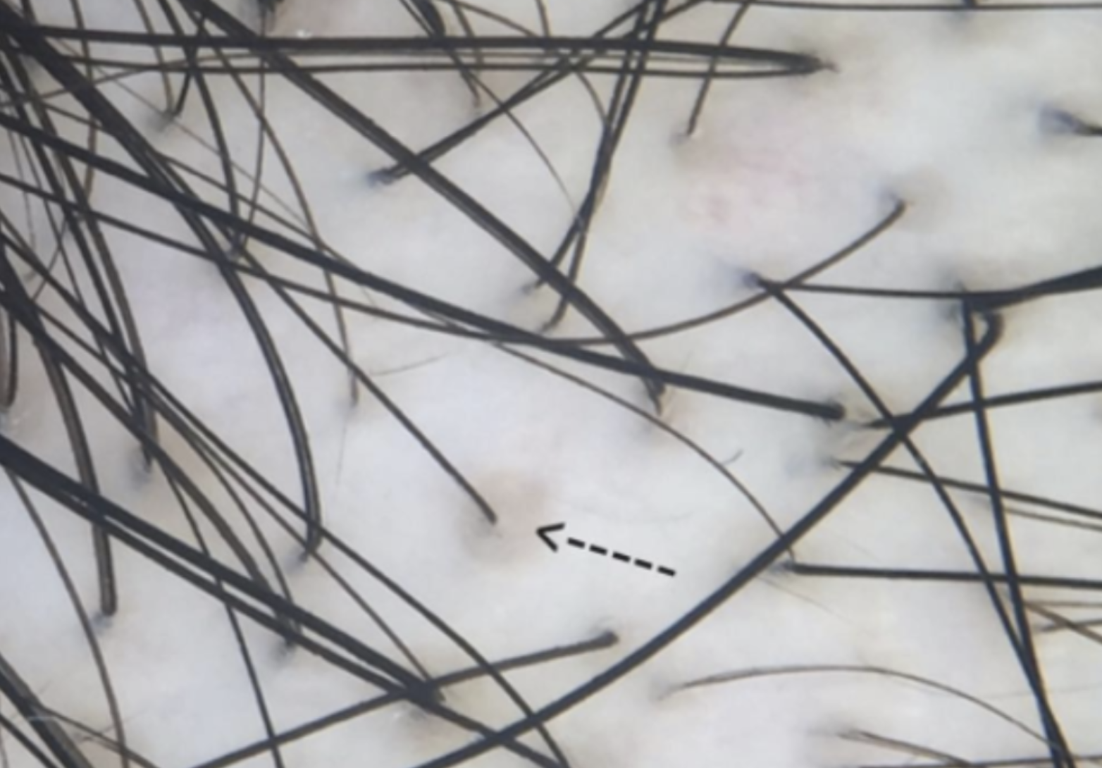
The brown peripilar sign (BPPS) was proposed as a trichoscopic sign in patients with AGA. The PPS is defined as the presence of a brown halo, roughly 1 mm in diameter, at the follicular ostium. It is thought to represent the inflammation beneath the scalp in patients with AGA. Let’s look at one of the original 2004 studies that led to the current understanding of the BPPS and then at a new study that calls this all into question!

Brown peripilar sign (arrow) in a patient with androgenetic alopecia (AGA)


The 2004 Deloche Study

In 2004, Deloche and colleagues from France studied 40 patients with androgenetic alopecia. The researchers showed that the brown discolouration around hairs seen with dermoscopy correlated nicely with inflammation under the scalp when evaluated by biopsy. They called this the peripilar sign (PPS) and proposed that it was an early sign of AGA. They found it present in 90% of males and 86% of females with AGA.

“Peri” means around, and pilar means hair. The peripilar sign is also known by many other names, including the “brown peripilar sign (BPPS)” and “peripilar halo.”


Abdalla D et al. 2024

Authors from Egypt set out to study the significance of the peripilar sign (PPS) in a cohort of patients with AGA and to evaluate whether it is associated with perifollicular inflammation.

The authors recruited 100 patients with trichoscopically confirmed AGA. The study group included 87 (87%) females and 13 (13%) males. Patient ages ranged from 16 to 67, and the duration of hair loss ranged from 0.16 to 20 years.

Peripilar signs were present in 50% of the 100 cases studied. Scalp biopsies were performed on 22 patients with PPS and 23 patients without PPS.

Interestingly, perifollicular inflammation on biopsy was present in AGA patients with PPS as well as in those without PPS. PFI was present in 82.2% of all studied biopsies. Inflammation was rated as mild in most patients (89.2%). The inflammation surrounded the infundibulum in 100% of cases. No significant difference was identified between those with and without PPS regarding the amount of inflammation.

The peripilar sign was more often encountered in patients with skin type III (p=0.001). Patients without the PPS had fewer yellow dots. Histopathologically, biopsies from patients with the peripilar sign were significantly associated with absent melanophages (p=0.011).


Comment

This is an interesting study that questions the meaning of the peripilar sign. The authors propose that the peripilar sign in AGA is not linked to perifollicular inflammation. They feel it is a dark colour in those with lighter skin types.

Further studies are needed to verify and extend our understanding of the BPPS.

The authors wonder whether UV radiation is somehow affecting the melanocytes.


REFERENCES*

Abdalla D et al. Peripilar Sign in Androgenetic Alopecia: Does It Really Indicate Peripilar Infiltrate? Dermatol Pract Concept . 2024 Jan 1;14(1):e2024096. doi: 10.5826/dpc.1401a9


Deloche C et al. Histological features of peripilar signs associated with androgenetic alopecia. Arch Dermatol Res. 2004.


This article was written by Dr. Jeff Donovan, a Canadian and US board certified dermatologist specializing exclusively in hair loss.
Share This
No Comments

New Study Adds Support to Notion that COVID19 Can Trigger Alopecia Areata

COVID-19 infection can trigger several autoimmune diseases, including rheumatoid arthritis, lupus, and many more. (For further review, see the article “Autoimmune Disease Risk Increased After COVID19 Infection.) There is growing evidence that COVID-19 infection can trigger AA or flare AA in those who already have the disease.

  • A 2023 study by Lim et al. showed an increase in both alopecia areata and alopecia totalis following COVID infection and a reduced risk in those who were vaccinated

  • A 2023 study by Tesch  et al. et al. showed an increase in both alopecia areata following COVID infection

Kim et al. 2024

A new study supports the notion that COVID-19 infection increases the risk of all forms of alopecia areata in adults.

The authors conducted a cohort study to investigate the association between COVID-19 and AA. They studied patients between October 8, 2020, and September 30, 2021.

The study comprised 259 369 patients with COVID-19 and 259 369 uninfected controls.

Results

To begin with, the authors found an increased risk of telogen effluvium in the cohort with COVID-19 compared with the uninfected cohort (AHR, 6.40; 95% CI, 4.92- 8.33).

Incidence of AA Increased in COVID-19.

The incidence of alopecia areata in patients with COVID-19 was found to be almost two times higher than in patients who did not have COVID infection (i.e. 43.19 per 10 000 person-years [PY] vs 23.61 per 10 000 PY)

Incidence of both Patchy AA and AT/AU Increased in COVID-19.


The authors studied the incidence of mild and severe alopecia areata separately. They showed that the incidence of all forms of alopecia areata increased with COVID-19 infection.

For patchy AA. The incidence of patchy AA was 35.94 per 10,000 PY in patients with COVID-19 and 19.43 per 10,000 PY in controls.

For AT/AU. The incidence of alopecia totalis and unversalis was 7.24 per 10,000 PY in patients with COVID-19 and 4.18 per 10,000 PY in controls.

Adult Patients and Females at Higher Risk

The incidence of AA after COVID-19 was significantly increased in all groups older than 20 years. In this study, the risk of AA was not increased in children and adolescents following COVID-19. There was a higher risk among both females and males, with a higher risk in females than males.


The Prevalence of Alopecia is Increasing in post-pandemic Korea

The authors noted that the incidence and prevalence of AA and AT/AU remained constant from 2006 to 2015. They point out that the prevalence has now increased following the COVID-19 pandemic.


REFERENCES**

Kim J-S et al. Risk of Alopecia Areata After COVID-19. JAMA Dermatol. 2024 Feb 1;160(2):232-235. doi: 10.1001/jamadermatol.2023.5559.

Christensen RE, Jafferany M. Association between alopecia areata and COVID-19: a systematic review.JAAD Int. 2022;7:57-61. doi:10.1016/j.jdin.2022. 02.002 3.

Birkett L, Singh P, Mosahebi A, Dhar S. Possible associations between alopecia areata and COVID-19 vaccination and infection. Aesthet Surg J. 2022; 42(11):NP699-NP702. doi:10.1093/asj/sjac165


Kutlu Ö, Aktaş H, İmren IG, Metin A. Short-term stress-related increasing cases of alopecia areata during the COVID-19 pandemic. J Dermatolog Treat. 2022;33(2):1177. doi:10.1080/09546634.2020.1782820


Kim J, Hong K, Gómez Gómez RE, Kim S, Chun BC. Lack of evidence of COVID-19 being a risk factor of alopecia areata: results of a national cohort study in South Korea. Front Med (Lausanne). 2021;8:758069. doi:10.3389/fmed. 2021.758069


This article was written by Dr. Jeff Donovan, a Canadian and US board certified dermatologist specializing exclusively in hair loss.
Share This
No Comments

Gut Microbiome Changes in Androgenetic Alopecia: The Good and the Bad

Specific Gut microbiota Alterations Linked to AGA

The gut microbiome refers to the array of organisms that live in our bowels. It’s clear that the health of the gut microbiome is extremely important for human overall health. An altered gut microbiome can lead to disease.

A recent study sought to identify changes in the microbiome associated with androgenetic alopecia and those felt to help prevent AGA.

Risk Factors for AGA included the genus Olsenella, genus Ruminococcaceae UCG-004, and genus Ruminococcaceae UCG-010

Preventive factors for AGA included the family Acidaminococcaceae and genus Anaerofilum, along with the genus Ruminiclostridium

Comment:

This was an interesting study. In the coming years, we’ll hear much more about our microbiome and how various changes influence human health.

REFERENCE*

Fu H et al. Roles of gut microbiota in androgenetic alopecia: insights from Mendelian randomization analysis. Front Microbiol. 2024 Apr 2:15:1360445. doi: 10.3389/fmicb.2024.1360445. eCollection 2024.


This article was written by Dr. Jeff Donovan, a Canadian and US board certified dermatologist specializing exclusively in hair loss.
Share This
No Comments

Does minoxidil cause hair greying? A new study suggests the answer is 'yes'

26% of Patients Felt Minoxidil Causing Greying

Authors from Saudi Arabia set out to evaluate whether minoxidil can affect hair greying. They used two surveys: one given to patients and one given to dermatologists.

Patient Survey

The patient survey comprised 453 patients. 56.7% of patients were aged 18-24 and mostly female. 26% of patients felt that minoxidil caused hair greying, and 14.8% noticed other colour changes (such as yellow, orange, and light brown). Hair colour changes occurred within the first five months in about one-third of women, but many had more delayed colour changes. Patients using minoxidil for a longer duration and patients with a family history of greying were more likely to report hair greying.

Dermatologist Survey

In the second surgery, 57 dermatologists completed the survey. 60% of dermatologists reported that they observed hair greying after minoxidil use. 17% of dermatologists who saw grey hair felt that minoxidil was responsible for the greying of hair.

Conclusion

Overall, the authors proposed that prolonged use of minoxidil can cause hair greying, especially in those with a family history of greying.

I really enjoyed reading this study! Every day, I’m faced with numerous questions from patients. A common question is, “Do you think minoxidil is responsible for my greying of hair?” This study suggests that it very well could be!

It’s interesting to me that the product monograph for Rogaine states, “some patients have experienced changes in hair colour and/or texture with ROGAINE MINOXIDIL TOPICAL 2% SOLUTION, Men’s ROGAINE FOAM 5% or Women’s ROGAINE FOAM 5% use.”

What’s even more interesting to me is that the product monograph errs on the side of extreme safety in advising patients to stop using the product if this occurs.

This is not always practical, as some patients will lose hair if they stop! What the product monograph should say if truly the company feels patients should stop is “We advise patients to stop using the product if hair colour changes occur and accept the fact that they may experience hair shedding and hair loss from stopping”

More studies are needed to better understand this phenomenon and determine whether the current era of extreme popularity of oral minoxidil use will lead to more people noticing grey hair.

REFERENCE*

Alhayaza G et al. Topical minoxidil reported hair discoloration: a cross-sectional study. Dermatol Reports. 2023 Aug 10;16(1):9745. doi: 10.4081/dr.2023.9745. eCollection 2024 Mar 12.


This article was written by Dr. Jeff Donovan, a Canadian and US board certified dermatologist specializing exclusively in hair loss.
Share This
No Comments

Topical Minoxidil: Not Great for Patchy AA, but not too Bad Either!

Topical Minoxidil Inferior to Betamethasone dipropionate in Treatment of Patchy Alopecia Areata

I enjoyed reading a study from Pakistan comparing topical betamethasone dipropionate and topical minoxidil in the treatment of patchy alopecia (less than 50% loess). This non-randomized study involved 100 patients—50 received topical betamethasone dipropionate twice daily, and 50 received topical minoxidil twice daily. The study lasted four weeks.


The authors showed that topical betamethasone dipropionate was superior to topical minoxidil in helping people regrow hair. 74 % of patients in the betamethasone dipropionate group achieved a 50 % or more improvement in their hair, compared to 42% of patients in the minoxidil-only group. However, the point that must not be missed in this study is that topical minoxidil still helped!


Conclusion

The study's conclusion is, of course, that topical betamethasone dipropionate twice daily is better than topical minoxidil 5% twice daily for patchy alopecia areata.

What I liked in this study is something that is ignored and pushed aside - that topical minoxidil still performed reasonably well! It was not as good as topical betamethasone dipropionate, but it had a respectable performance.

Let’s face it—topical betamethasone dipropionate and topical minoxidil are both pretty safe over a short period, like four weeks. But you can’t keep rubbing on betamethasone dipropionate forever. Prolonged use can thin the skin and even contribute to side effects if too much is used. One must take breaks every now and then if betamethasone dipropionate is going to be used.

Topical minoxidil, on the other hand, is reasonably safe for continued use. It does not thin the skin, and it is widely available and does not require a prescription in many countries.

One must always remember that topical minoxidil still has a role in the alopecia areata treatment toolbox!


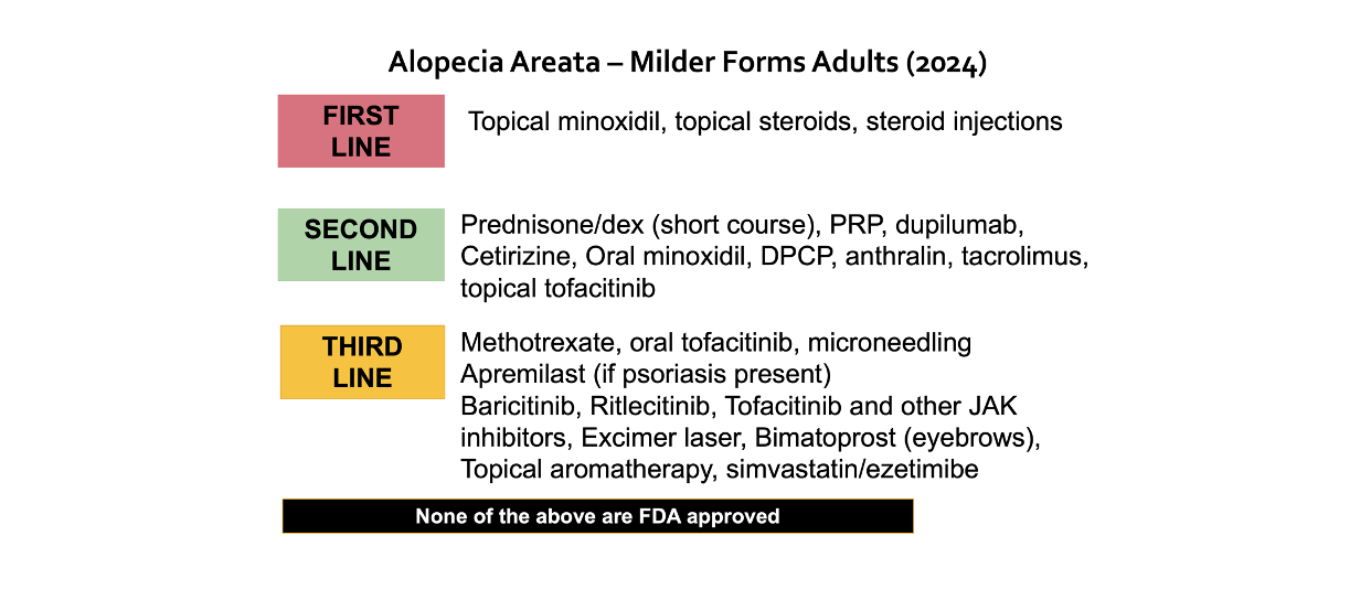
Here are my first line, second line and third line options for the treatment of alopecia areata


REFERENCES

Aslam S et al. Comparison of Efficacy of Topical Betamethasone Dipropionate and Topical Minoxidil in Patients With Alopecia Areata. Cureus . 2024 Mar 16;16(3):e56282. doi: 10.7759/cureus.56282. eCollection 2024 Mar.


This article was written by Dr. Jeff Donovan, a Canadian and US board certified dermatologist specializing exclusively in hair loss.
Share This
No Comments

The Benefits of Topical Minoxidil for Facial hair

Topical Minoxidil helps Facial hair Growth

Many males value good facial hair growth. Growth patterns are strongly genetically determined. There is a tremendous amount of information online about using topical minoxidil to enhance facial hair. Despite all these anecdotal reports, few studies have been done. An important study to note was a 2016 study by Ingprasert et al. This was a randomized study of 48 males using 3 % minoxidil for facial hair growth (…to review, click on the link).


Shokravi and  Zargham, 2024

In a new study, the authors present a case showing the potential benefits of topical minoxidil for facial hair. What was unique in this report was that the patients were identical twin males. One used minoxidil, and one did not. The twins had similar facial hair density before using minoxidil.


TWIN 1: used ¾ cap topical 5% minoxidil foam for over a year on the beard and mustache area

TWIN 2: did not use topical minoxidil


Results in Treated Twin

The study's authors described that the twin using minoxidil had improvements in facial hair that were not seen in the untreated twin. New, finer, lighter-colored hairs were noted by month 1. A modest increase in facial hair density was noted by month 2. The treated twin reported shedding of the hairs in the beard area approximately three months after initiating minoxidil.

Overall, after 16 months of use, the minoxidil-treated twin had a greater hair count and density in both the beard and mustache areas.


Photos show the differences in facial hair ten days after shaving.

SIDE EFFECTS

There were no major side effects.

Minimal mild side effects with foam minoxidil use included:

  • Mild skin dryness

  • hypertrichosis on the ears and forehead

  • An increase in body hair (chest, abdomen, forearms, and legs)


Interestingly, the patient initially used minoxidil solution but switched to the foam due to DRY FLAKY SKIN.


CONCLUSION

This study concluded that topical minoxidil is one strategy to consider for males wishing to improve facial hair growth.


REFERENCES*

Shokravi A and  Zargham  H. Facial hair enhancement with minoxidil-an off-label use. SAGE Open Med Case Rep. 2024 Feb 23:12:2050313X241231490. doi: 10.1177/2050313X241231490. eCollection 2024.

Ingprasert S et al. Efficacy and safety of minoxidil 3% lotion for beard enhancement: A randomized, double-masked, placebo-controlled study. J Dermatol . 2016 Aug;43(8):968-9. doi: 10.1111/1346-8138.13312. Epub 2016 Feb 19.


This article was written by Dr. Jeff Donovan, a Canadian and US board certified dermatologist specializing exclusively in hair loss.
Share This
No Comments

Patients Using Dupilumab Have an Increased Risk of Being Diagnosed with Cutaneous T Cell Lymphoma More Often

Dupilumab and CTCL: What does it mean for our alopecia areata patients?


I’ve been increasingly prescribing dupilumab, the IL4/13 antagonist, in the management of some patients with alopecia areata and atopic dermatitis. Dupilumab is a drug often used for atopic dermatitis but has the potential to help hair growth in a small proportion of patients with alopecia areata. Safety seems reasonably good. Blood tests are not needed for most, and it’s reasonably easy to administer.

We’ve talked about the role of dupilumab in alopecia areata in prior articles.

But then there’s the lymphoma studies. Studies keep coming out showing a similar trend: if you are prescribed dupilumab, you have an increased chance you’ll be diagnosed with a specific skin lymphoma known as cutaneous T cell lymphoma. It’s not a massively increased risk, but it’s an increased risk.

I’ll review a new study in a moment, but let’s talk about this subject!

Challenges in the CTCL Data

Let’s face it; the data is challenging to decipher. On the one hand, some feel the lymphomas associated with dupilumab are not really lymphomas at all. Boesjes CM et al., in 2023, showed that dupilumab treatment can cause a reversible and benign ‘lymphoid reaction’ that mimics a CTCL but is not CTCL.

On the other hand, there are more and more and more reports showing a massive “unmasking” of CTCL following the initiation of dupulimab therapy. The cases showing that some of the patients treated with dupilumab end up dying are a stark reminder of how important this issue is.

Espinosa et al. 2020

In 2020, Espinosa et al. described four patients given dupilumab for presumed atopic dermatitis and 3 for use off-label to treat pruritic cutaneous T-cell lymphoma. (In cutaneous T-cell lymphoma, IL-13 is overexpressed and regulates cell proliferation, so it was initially felt it could be helpful to block tumour progression.)

All patients treated with dupilumab had disease progression. The four patients initially presumed to have atopic dermatitis subsequently received a diagnosis of cutaneous T-cell lymphoma. The three patients with existing cutaneous T-cell lymphoma progressed to Sézary syndrome while receiving treatment, and two died.

Jfri A et al. 2023

Jfri et al. systematically reviewed twenty-three dupilumab-associated CTCL cases from 11 sites.

The median (range) age was 58 (27-74). Atopic dermatitis onset was mostly in adulthood in 19 (82.61%) cases and childhood-onset in 4 (17.39%) cases. In 13 patients (56.52%), skin biopsies done before starting dupilumab were not diagnostic of MF/SS. In 5 patients, there were atypical features for AD (epidermotropism, folliculotropism, lichenoid inflammation).

The mean time between the start of dupilumab and biopsy-proven cutaneous T-cell lymphoma was 7.5 months. Eleven had advanced disease (Stage IIB-IV) at diagnosis. Personal/family atopic history, a major criterion for clinical AD diagnosis, was found in only 4 of 23 cases. Several patients initially diagnosed with atopic dermatitis had features that are not typical of AD, including indurated plaques, nodules, poikiloderma, and sparing of flexural sites.

When looking back at these cases, the authors found that there were two cases where it was clear that the patient had atopic dermatitis to begin with. These are 2 cases with a long-standing diagnosis of AD since childhood, and biopsies done before starting dupilumab showed features consistent with AD. Of those 19 patients with adult-onset atopic dermatitis, only five patients had clinical presentations and pre-dupilumab biopsies truly consistent with AD. In many cases, it was really not possible to exclude that patients had skin lymphoma before they started therapy.

Hasan et al. 2024

In a new study, Hasan and colleagues used a large database to compare the incidence of CTCL in those who used dupilumab compared to those who never used dupilumab.

This was an extensive database. Data was extracted from 60 healthcare organizations, encompassing 22,888 AD patients who were prescribed dupilumab and 1,115,235 AD patients who were not prescribed dupilumab,

The cohort of AD patients who used dupilumab had an increased risk of CTCL (OR 4.1003, 95% CI 2.055-8.192). The risk was not increased for other cutaneous or lymphoid malignancies, like non-Hodgkin lymphoma or leukemia. Most (27/41) cases of CTCL were diagnosed more than one year after dupilumab use, suggesting that perhaps these are not simply unmasked cases.

So does dupilumab Cause or UnMASK Skin Lymphoma?

It’s pretty clear that if you use dupulimab for atopic dermatitis, you are at increased risk of being diagnosed with a type of skin rash that causes everyone to worry. That is something that is undebatable. It’s also clear that if you have a skin rash before starting dupilumab and it does not respond to treatment with dupilumab or gets worse, it needs to be biopsied to make sure one is not missing a diagnosis of CTCL.

We don’t yet have evidence that patients who use dupilumab for alopecia areata who do not have atopic dermatitis develop skin lymphoma or lymphoid reactions.

What is debatable is whether dupilumab actually causes a lymphoma or lymphoid-like reaction or whether patients had this skin malignancy to begin with, and dupilumab simply encouraged it to come out. Atopic dermatitis and cutaneous T-cell lymphoma can certainly mimic each other, and a widely held concern is that some of the cases of CTCL with dupilumab were simply cases that were first misdiagnosed as atopic dermatitis.

Most studies in the literature conclude their study with the unsatisfying sentence that goes something like “These data raise the question as to whether the affected patients were first misdiagnosed as having atopic dermatitis and the CTCL was unmasked by dupilumab or whether cutaneous T cell lymphoma is truly is an adverse effect of treatment with dupilumab. Other studies end with comments like lymphomas developing with dupilumab might be unrelated to the drug use itself, but long-term follow-up data of large patient cohorts is necessary to rule out such a possibility. Even the discussion section of the paper by Hasan et al. starts out with, “It is unknown if dupilumab causes CTCL or unmasks CTCL.”

For now, it’s clear that patients with atopic dermatitis who are considering dupilumab need one (or more) skin biopsies

  1. Their atopic dermatitis starts in adulthood (instead of childhood)

  2. They have a diagnosis of atopic dermatitis but don’t have a strong family history

  3. They have significant body surface area (erythroderma) - also need flow cytometry

  4. They don’t have involvement of the flexures

  5. They have atypical features like nodules or poikiloderma

Patients who start dupilumab and develop new rashes or worsening of existing rashes also need a low threshold to have biopsies.

I’m following this field closely!


REFERENCES*

Hasan I et al. Dupilumab therapy for atopic dermatitis is associated with increased risk of cutaneous T cell lymphoma: a retrospective cohort study. J Am Acad Dermatol. 2024 Apr 6:S0190-9622(24)00566-8. doi: 10.1016/j.jaad.2024.03.039. Online ahead of print.

Boesjes CM et al. Dupilumab-Associated Lymphoid Reactions in Patients With Atopic Dermatitis. JAMA Dermatol . 2023 Nov 1;159(11):1240-1247.


Espinosa ML et al. Progression of cutaneous T-cell lymphoma after dupilumab: Case review of 7 patients. J Am Acad Dermatol . 2020 Jul;83(1):197-199. doi: 10.1016/j.jaad.2020.03.050. Epub 2020 Mar 27.


Harada K et al. The effectiveness of dupilumab in patients with alopecia areata who have atopic dermatitis: a case series of seven patients. Br J Dermatol. 2020 Aug;183(2):396-397.


Poyner EFM et al. Dupilumab unmasking cutaneous T-cell lymphoma: report of a fatal case. Clin Exp Dermatol. 2022 May;47(5):974-976. doi: 10.1111/ced.15079. Epub 2022 Feb 9.

Du-Thanh A et al. Lethal anaplastic large-cell lymphoma occurring in a patient treated with dupilumab. JAAD Case Rep. 2021 Oct 12:18:4-7. doi: 10.1016/j.jdcr.2021.09.020. eCollection 2021 Dec.

Jfri A et al. Diagnosis of mycosis fungoides or Sézary syndrome after dupilumab use: A systematic review. J Am Acad Dermatol . 2023 May;88(5):1164-1166. doi: 10.1016/j.jaad.2022.12.001. Epub 2022 Dec 5.


This article was written by Dr. Jeff Donovan, a Canadian and US board certified dermatologist specializing exclusively in hair loss.
Share This
No Comments

Frontal Fibrosing Alopecia in India: A Reminder of a Truly World-Wide Epidemic

First Report of FFA Cases in North-East India

Several months ago, I had a discussion with a several colleagues. We spoke about FFA and the role of stress, diet, cosmetics, family history, hormones. Basically … everything!

Not too long into the discussion, a point was made by my colleague: “I don’t think we’re seeing much FFA in India!” Another made the point that if this is indeed true that there must be important dietary and genetic reasons that need exploration.

A new report in the Indian Journal of Dermatology reminds us that while FFA in India might be a little less frequent than some pockets of the world, it’s probably understudied - and by no means absent from India!

Authors from North-East India, in Meghalaya state, reported 21 patients with FFA that they saw at their center over the period 2013-2023. 19 were female and the average age was 48 years. 28% had lichen planus pigmentosus of the skin. Only 10 % had androgenetic alopecia and only 10 % had classic LPP.


Comment

This was interesting to see. In this small world we live in with global travel and global products and expanding ingredients, pollution, cosmetics, etc, one can only imagine that FFA must be increasing in most part of the world.

More studies are needed but I congratulate these authors!


REFERENCE

Verma S et al. A Retrospective Study of Frontal Fibrosing Alopecia from North-East India. Indian J Dermatol. 2023 Nov-Dec;68(6):598-602. doi: 10.4103/ijd.IJD_290_23. Epub 2024 Jan 9.


This article was written by Dr. Jeff Donovan, a Canadian and US board certified dermatologist specializing exclusively in hair loss.
Share This
No Comments

1 in 20 Drugs that Get Approved Will Eventually Be Removed From Market

When a new drug comes to market, I’m pretty excited. After all, it’s proof of something I fundamentally believe in : good science can create better treatments and help more people lead fuller lives.

Whenever a new drug comes to market, I also remember something I learned many years ago: about 5 % of the time, the drug you are cheering about is going to eventually be removed from the market because of the harm it caused someone.


Why are drugs removed?

Drugs are removed from the market for several reasons. The top 4 reasons seem to be hepatotoxicity, cardiac disorders hypersensitivity and kidney disease nephrotoxicity. Of course, these are not the only reasons.

According to Craveiro et al, it tends to be the case report and spontaneous reporting that triggers the steps that ultimately lead to removal of a drug - rather than some piece of evidence from a well designed clinical trial. This is important for all of us to remember. Yes, completing that side effect reporting form does make a difference.


How long does it take to get a drug off the market?

A study in Canada by Lechin found that if a drug is going to be removed from the market, then 50% will be gone by 3.5 years and 50% will stay on longer than 3.5 years. A 2020 study by Craveiro NS et al. showed the delay could be even longer. In their study, the average time (rather than median) between the introduction of a drug and its withdrawal due to safety reasons was 20.3 years (SD±13.8).


Comment

Yes, some of the drugs we prescribe are going to eventually be found to harm people seriously enough that they get removed from the market. I’m hoping that number drops lower and lower and lower below 5 %. I still remember the day in 2009 that I learned that Raptiva was removed from the market. I still remember the day in 2009 that I learned that a drug I had prescribed (Raptiva) for psoriasis was removed from the market. The FDA approval of Raptiva was based on initial studies in approximately 2,700 patients - most received the drug for only a few months. Fast forward 6 years to 2009, after over 46,000 patients had received efalizumab. The world came to learn that 3-4 patients developed a rare and serious disease known as progressive multifocal leukoencephalopathy (PML) after using Efalizumab. 3 died. The only way that these side effects came to be realized was through reporting of side effects “after” the drug was released: doctors and patients saw the side effect and reported it regardless of whether they felt it was truly linked or not. Over time, it became clear it was linked.


The next 20 years will see a tremendous number of new treatments in our field. It’s going to be incredible. I’m exited to be part of this expansion of options for our patients.


But approximately 5 % of the time, we’re going to have it all wrong.


REFERENCE

Joel Lexchin. How safe are new drugs? Market withdrawal of drugs approved in Canada between 1990 and 2009. Open Med. 2014; 8(1): e14–e19. Published online 2014 Jan 28.

Craveiro NS et al. Drug Withdrawal Due to Safety: A Review of the Data Supporting Withdrawal Decision. . Curr Drug Saf. 2020;15(1):4-12. doi: 10.2174/1574886314666191004092520.


This article was written by Dr. Jeff Donovan, a Canadian and US board certified dermatologist specializing exclusively in hair loss.
Share This
No Comments


Share This
-->